Each neuron also sends
out dendrites that
will eventually let it receive contacts from other neurons.
Dendrites are extensions from the cell body that are shorter
than axons and are highly ramified (branched). Like axons,
dendrites grow by extending growth cones at their tips.
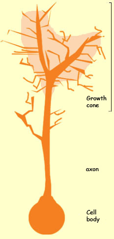
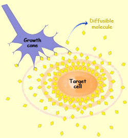
THE GROWTH CONE
Probably one of
the most amazing things about the way the nervous system
develops is how the growing axons find their target cells,
even though these cells are often located millimetres
or even centimetres away (a vast distance on this scale).
The source of this ability is the growth cone,
a structure at the tip of each elongating axon.
The growth ’cone is composed of various
projections that extend and retract as they seek out
signals to guide them, something like your fingers might
if you were groping to explore your surroundings in the
dark. By interacting in this way with its environment,
each growth cone finds signals that guide it to the spot
where it must establish
connections with its target cells .
These guidance signals consist of molecules
that tell the growth cone which direction to go.
Some of these guidance molecules are attached directly
to the substrate along which the growth cone moves.
But other guidance molecules are secreted by cells
and diffuse freely into the surrounding area. A concentration
gradient thereby forms that remotely influences the
path that the axon follows as it grows.
The reason that the growth cone can be
influenced by these molecules is that it has special receptors to
detect them. Thus it is through the deployment of guidance molecules
and the distribution of specific receptors for them across various
neurons that the major neural pathways are laid down in the embryo.
There are over 100 billion
neurons in the human brain, and each of them forms several
thousand synapses with other neurons. The possible combinations
thus greatly exceed the 20 000 or 30 000 genes
in the human genome. This limited nature of the available
genetic information suggests that other, extrinsic factors,
such as chemoattractive molecules and interactions among
cells, play a major role in the development of the nervous
system.
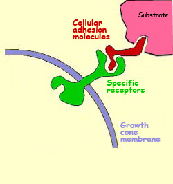
THE MOLECULES THAT GUIDE THE GROWTH CONE
The growth cone that guides
the axon to a cell with which it must form
a synapse is like someone driving a car through unfamiliar country
with no road map and only the signs along the way as a guide. For
the growth cone, these road signs take the form of molecules. These
growth cone guidance molecules are divided into two major families.
The
first family consists of molecules that are attached
to various substrates along the path that the growth
cone travels. Like signs that a driver recognizes alongside
the road, these cell adhesion molecules are
recognized by specific
receptors on the growth cone’s membrane.
As the result of direct contact between the cell
adhesion molecules and their receptors on the axon’s
growth cone, other signals are transmitted to the
inside of the growing axon. These signals ultimately
set the direction in which the axon grows. In contrast
to the other family of guidance molecules, cell adhesion
molecules are described as “non-diffusible”.
Source:
Dr. Brian E. Staveley
Department of Biology
Memorial University of Newfoundland
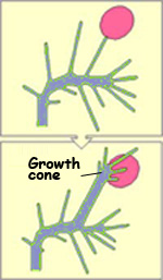
Growth cone changing direction after touching a substrate
with compatible cell adhesion molecules
The second family of axon guidance
molecules are not attached to a substrate. Instead, they
diffuse freely into the aqueous environment surrounding
the growth cone. The mechanism by which these diffusible
molecules guide the growth cone is called chemotropism.
Some of these molecules guide the growth cone by attracting
it, the way the smell of freshly brewed coffee pulls
a coffee lover toward the shop where it is being brewed.
This kind of chemotropism is called “chemoattraction”.
But there are also some guidance molecules that repel
the growing axon, just as foul odours from a landfill
might repel someone walking by. This kind of chemotropism
is called “chemorepulsion”.
Lastly, there is a third category of molecules that do not
provide guidance signals as such but are nevertheless necessary
for the elongation of the axon. These molecules are called growth
factors, and they play a crucial role in the formation
of synaptic connections.
In addition to allowing certain
neurons to survive during development and
then participate in organizing their initial connections,
the competition for trophic molecules allows the neurons’ ramifications
and connections to change throughout the individual’s
lifetime, in response to changes in neural activity
caused by learning
processes.
TROPHIC FACTORS AND NEURONAL DEATH
From the moment that the neurons start
to form circuits, a change of scale takes place in the nervous
system’s development—from
isolated cells to a network of thousands of interconnected
elements. It is this network that develops the information-processing
capacity that makes the brain such a powerful tool.
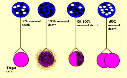
But initially, this network is far from perfect. At first, the embryo produces
two to three times more neurons than it needs. Subsequently, the excess neurons
die off. So which neurons survive, and why?
We now know that a neuron’s survival depends largely
on the relationship that it maintains with its target cell.
For example, experiments have shown that reducing the number
of target cells reduces the number of neurons that have to
come connect to them. Conversely, the existence of a larger
population of cells that have to be innervated keeps a larger
number of innervating neurons alive.
The survival rate of neurons depends on
the size of the population of target cells that they innervate.
The shaded areas here represent the destruction of target cells.
The mechanism at work here involves a certain competition among
the neurons for special substances known as growth factors or trophic
factors (from the Greek trephein, meaning “to
nourish”). These factors are not energy sources like glucose
or ATP. But they are molecules
that are secreted by the target cells and that the neurons
must have in order to survive.
To use an automobile metaphor again,
these factors are not the gasoline that the car uses as its energy
source; they are more like the driver’s licence. The absence
of growth factor leads to major problems in the development of
an axon and very often to its outright removal from the neural
highway.
The way neurons die in such circumstances is quite different from
their death due to injury or illness. Instead it is a gradual,
programmed form of death, known as apoptosis.
Apoptosis involves the expression of a multitude of specific genes
that make cells decay in a way that does not harm the organism
as a whole. These same genes are also often involved in cell differentiation
and in controlling the normal cell life cycle. (For more about
apoptosis, follow the Tool module link at the start of this section.)
The neurons in a baby’s
brain receive about one and a half times more synapses
than those in an adult’s. It is thought that the
number of these myriad connections remains fairly constant
until puberty, but declines markedly during adolescence.
For example, during adolescence, the neurons of the primary
visual cortex are believed to lose an average of 5 000
synapses per second!
The process whereby
a target cell loses connections from several neurons and
retains connections from only one is often inaccurately
referred to as synapses’ being eliminated. It would
be more accurate to say that there is a reduction in the
number of different afferences that the target cell receives.
The total number of synapses generally does nothing but
increase over the course of development.
FORMATION AND SELECTIVE STABILIZATION OF SYNAPSES
Often, the axons of neurons that will ultimately be part of
different neural circuits are guided along
the same path until they reach the vicinity of their target
cells. But how does each axon then recognize its own target
cell?
In some cases, molecules similar to cell
adhesion molecules seem to act as labels that let the
various axon growth cones recognize
the right target cells. In addition, the exact location where
the axon forms its synapse with
the cell is closely controlled by a particular set of molecules,
because the synapse requires the presence of particular molecular
structures in order to function properly.
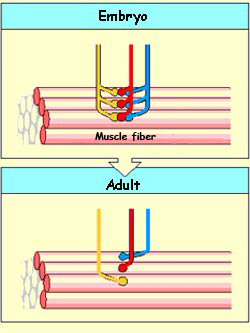
In a neuromuscular
junction (the type of synapse whose formation
process has been studied the most), this location
is called the “active zone”. Here the
axon’s machinery for releasing neurotransmitters
from the motor neuron aligns with very dense groups
of acetylcholine
receptors in the muscle fibre.
Initially, one muscle fibre may receive connections
from several motor neurons. But gradually, it will
lose all but one of these junctions and remain connected
to only one motor neuron.
Researchers have shown that this process is regulated by the
electrical activity in the muscle fibre. The more active the
fibre, the more quickly synapses are eliminated, except for
those from the one motor neuron that will remain . Conversely,
reducing the muscle fibre’s activity slows down this
selection process.
There is ample evidence that such synaptic reorganizations also occur in the
immature brain. A given neuron may lose connections that it had initially established
with other neurons, or it may see its connections with certain neurons multiply.
Here again, it is neural activity that maintains or increases the number of synaptic
contacts, while the absence of stimulation leads to the elimination of synapses
that are unneeded. That is why the
stabilization of the synapses is regarded as a selective process and why
the activity of the neural circuits is regarded as controlling this selection.
The connections
among the neurons of the human brain are initially determined
by an inherited genetic map. Subsequently, these connections
are remodelled through the individual’s interactions
with the environment. Two major types of information
pathways then develop: converging pathways,
in which multiple nerve fibres connect to the same target
cell, and diverging pathways, in which
a single neuron connects to several different cells.