|
|
|
|
 |
From Embryo to Ethics |
|
|
|
|

The precursor
cells of neurons proliferate in a part of the neural tube
called the ventricular zone, adjacent to
the neural tube’s central canal. During the period
of maximum proliferation while the embryo is gestating, an
estimated 250 000 new neurons form in the ventricular
zone every minute! |
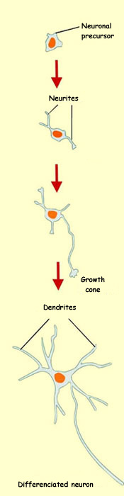
At a certain point in
its development, each stem cell that is destined to produce
a neuron divides into one stem cell and one neuronal precursor
cell, called a neuroblast. This neuroblast
continues to divide while forming various lineages of neurons.
At some point in time, the differentiated neuronal precursor
cell ceases the cycle of mitotic cell division. This moment
marks the birth of the resulting neuron.

|
Complex structures in
the nervous system, such as the spinal
cord and the cerebral
cortex, contain not just one but many
types of neurons. These various types of neurons
do not all develop at the same time. Often one type arises
from another, forming what are known as cell lineages.
One stem cell can thus give rise to various types of neurons
(sensory neurons and motor neurons, for example), often using
different neurotransmitters. |
The term neural
stem cells refers to all of the cells that are
the source of the various
types of neurons found in the brain and even
of the various
kinds of glial cells in the nervous system.
The ultimate fate of the migrating daughter cells therefore
does not depend on what kind of stem cell they come from,
but rather on a multitude of other factors, such as the
age of the precursor cell, its environment, and the orientation
of its cleavage plane when it divides. |
|
|
| HOW STEM CELLS FORM NEURONS |
|
How can a single cell, the
fertilized ovum (egg), give rise to so many different kinds of
cells in the human body, ranging from neurons to blood cells to skin
cells?
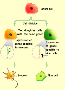
| To begin answering
this question in very simple terms, we should first remember
that every cell in the human body contains all of the genes
capable of forming a human being. The same is true of the cells
in a developing human body, especially if you go back to the
very start of an embryo’s life. A given cell acquires
its specific personality because only certain genes among all
those it possesses will be activated—the genes specific
to neurons or skin cells, for example. |
 |
The next question is: how do all the many different kinds of nerve
cells develop, and how do they manage to make their billions of
connections correctly in the human brain?
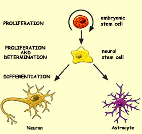
To attempt to answer this difficult question of the origin of our nerve cells,
we must look at three processes that act together to transform the precursor
cells, known as stem cells, into mature neurons.
The first of these processes is cell proliferation, which increases
the number of cells. Since the body needs to manufacture an astronomical number
of neurons—100 billion in the adult human brain—it has to start doing
so early in the embryo’s development.
Proliferation begins as soon as the closing
of the neural tube is completed. At this stage, the neural tube consists
of only a single layer of epithelial cells. But as soon as these cells
start to proliferate, this layer thickens rapidly (see sidebar).
|
The next process involved in
the origin of our nerve cells is called determination.
This is a critical stage in which the destiny of certain
cells is decided. They become the precursors that will eventually
give rise to various types of neurons and glial
cells.
As soon as the major
axes of the nervous system have been laid down, the
cells in each region can begin to differentiate.
Differentiation is the third process in the
maturation of the neurons. |
Through differentiation, a given population of neurons gives rise
to subpopulations that are specific to the various parts of the
nervous system. During this stage, the neurons continue to proliferate
and migrate
to their final locations, where they will make specific
connections with other neurons.
The cerebral cortex
is organized into numerous
columns that constitute the brain’s basic
information-processing units. The
expansion of the cortex that has occurred throughout the
course of evolution has been attributable to an
increase in the number of these columns, not in their individual
size.
Because each column originates in a small number of adjacent
stem cells, an increase in the number of columns must presumably
be due to an increase in the number of stem cells. It is
therefore highly likely that some minor changes in the
initial quantity of stem cells could be the source of the
vast cortical surface found in the brains of primates in
general and human beings in particular. |
|
|