| CORTICAL ATROPHY IN ALZHEIMER’S |
|
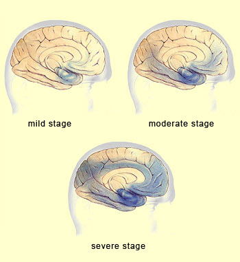
The areas shaded in dark blue in the above drawings show how cortical atrophy progresses over these three main stages of Alzheimer’s. This process is predictable as regards the sequence in which various cortical structures will be affected.
This predictability is due mainly to the way that neurofibrillary tangles proliferate; the build-up of amyloid plaques is a far more diffuse process.
The first areas of the cortex where atrophy is observed are those surrounding the hippocampus:
first the transentorhinal cortex, then the entorhinal cortex, and then the hippocampus itself. These structures play a key role in memory, so it is no coincidence that the first symptoms of Alzheimer’s
consist of memory deficits.
Interestingly, in most normal persons age 75 or older, the hippocampal area shows neurofibrillary degeneration comparable to that seen in the first stage of Alzheimer’s.
As Alzheimer’s progresses to the moderate stage, the atrophy gradually extends into the temporal cortex and into the prefrontal association cortex and the parietal-temporal-occipital association cortex.
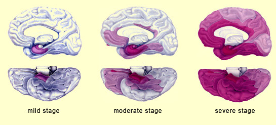
In the advanced stage of Alzheimer’s, the cortex as a whole is affected. The only parts that are sometimes spared are the primary visual cortex in the occipital lobe and the primary motor cortex.
Technologies such as brain imaging have enabled scientists to provide a more precise anatomical description of the stages of progression of Alzheimer’s disease and to show that neuronal death extends into various areas of the cortex along the connections between them.
|