|


Recent
Studies on the Role of Sleep
The discovery of an activation
of the hippocampus during REM sleep has lent
support to the idea that REM sleep plays a role in learning.
Many experiments have shown that we retain newly acquired
knowledge or a newly learned skill more effectively the
day after a good night’s sleep. And because the
hippocampus is known to be heavily involved in encoding
memories, REM sleep may thus contribute to learning
and memory.
The activity of the inferior parietal lobule,
a part of the cortex that conveys experiences to memory,
decreases during REM sleep, which probably helps to explain
why we have so much trouble in remembering our dreams. |
| |
| THE
BRAIN DURING REM SLEEP |
|
Electroencephalography is
a method of recording the activity of the cortex by
means of electrodes placed on the scalp. In the 1950s, electroencephalography
revealed that the cortex is just as active when someone is
in REM sleep as when he or she is awake. Scientists hence began
referring to REM sleep as “paradoxical sleep”,
to call attention to this phenomenon.
But with the development of brain imaging technologies in the mid-1990s (follow
the Tool module link to the left), researchers discovered other brain structures,
many of them located deep below the cortex, whose activity was greatly altered
during REM sleep. In some of these areas, activity increased during REM sleep,
while in others, it decreased. But what was remarkable was that this increase
or decrease in activity was consistent with the particular
kind of dreaming that occurs during REM sleep.
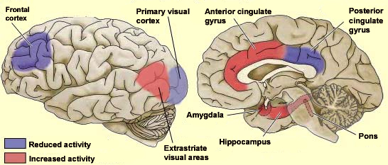
Brain imaging studies have found, for example, that the primary
visual cortex, the first part of the brain involved
in consciously decoding visual signals when people are awake,
shows very little activity when they are dreaming during
REM sleep. This is no surprise—when people are dreaming,
their eyes are closed, and no visual signals are reaching
them.
But brain imaging studies have also shown
that certain extrastriate
visual areasof the cortex, which decode complex
visual scenes, are significantly more active during REM sleep.
Thus, during REM sleep, these areas are apparently involved
in analyzing complex visual scenes. This is completely consistent
with the often highly elaborate visual dream scenes that
people report when researchers awaken them from REM sleep.

Adapted from Neuroscience,
Purves et al., from Hobson et al., 1998
During REM sleep, intense activity is also observed in the limbic
system, a set of structures heavily involved in
emotions. Two of these structures are especially active: the hippocampal
regionand, in particular, the amygdala. Once
again, it is interesting to note that this intense limbic activity
does not occur during the phases of non-REM
sleep , when the dreams that people have are far less emotional.
The frontal cortex is a part of the brain
that maintains very close ties with the limbic system. Yet the
frontal cortex remains relatively calm during REM sleep. The prefrontal
cortex, which is part of the frontal cortex, is heavily
involved in thought and judgment when we are awake. Its low activity
during REM sleep might thus account for the bizarre, illogical,
and often socially inappropriate content of people’s dreams.
The anterior cingulate gyrus, which governs
attention and motivation, is also more active during REM sleep,
which may be part of the reason that the images we see in dreams
are so vivid and so changeable.
Lastly, the pons is also
more active during REM sleep, which makes complete sense, because
even though the elaborate dreams that occur during REM sleep
certainly involve the cerebral cortex, it does not seem to be
involved in triggering REM sleep: REM
sleep is triggered by certain nuclei (groups of neurons) in the
pons.
| |


