Tool Module: Brodmann's Cortical Areas
In the brains of all vertebrate animals, the nerve cells of the cortex are characteristically organized into parallel layers at the brain's surface. At least one of these layers comprises pyramidal neurons with long apical dendrites that extend upward to the layer closest to the surface. This layer generally contains no cell bodies and consists chiefly of nerve fibres, dendrites and axons.

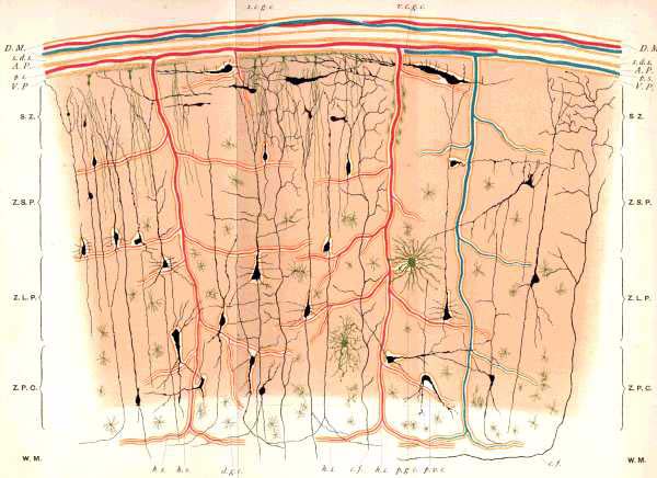
Golgi's stain, developed in the
early 20th century, was one of the first methods that enabled scientists to view
the neurons of the cortex.
Source: J. Batty Tuke, The Insanity of
Over-Exertion of the Brain, Edinburgh, 1894.
Some
parts of the cortex that are of very ancient origin, such as the hippocampus,
contain only a single layer of cells. Others, such as the olfactory cortex, contain
two layers. But the most elaborate cortex, the neocortex, contains at least six
well defined main layers, which are often divided into sublayers. In the course
of evolution, the neocortex appeared only with the mammals. In humans and other
primates, the neocortex is so dominant that the term "cortex" is often
used to refer to the neocortex alone.
The cellular architecture differs
sufficiently from one part of the neocortex to another to be used as a criterion
for defining cortical areas that are functionally distinct. That is what the German
anatomist Korbinian Brodmann did in the early 20th century, when he developed
a map of the brain based on the differences in the cellular architecture of the
various parts of the cortex. Brodmann assigned each part of the cortex that had
the same cellular architecture a number from 1 to 52.
Brodmann's intuition,
whose accuracy has been confirmed many times since, was that a particular anatomical
structure corresponded to a particular function.
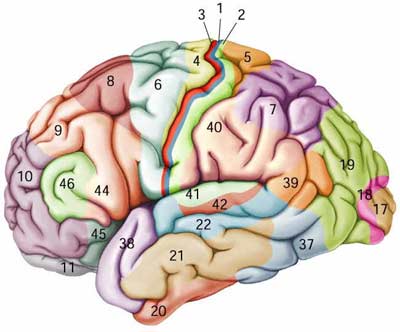
For example, Brodmann's
area 17, which receives information from a nucleus of the thalamus that is connected
to the retina, turns out to correspond precisely to the primary visual cortex.
And Brodmann's area 4, from which the axons of the large pyramidal cells project
to the motor neurons of the spinal cord, corresponds broadly to the motor cortex.

|
