Funding for this site is provided by readers like you.
Body movement and the brain
Sub-Topics
Making a Voluntary Movement

Linked
HelpLink : The Physiology of Neuromuscular TransmissionLink : Récepteur nicotiniqueLink : The mechanism of filament sliding during contraction of a myofibril
Link : LE MUSCLE STRIÉ SQUELETTIQUE - ORGANE EFFECTEUR DE LA MOTRICITÉ SOMATIQUELink : The Neuromuscular JunctionLink : MÉCANISMES DE LA CONTRACTION MUSCULAIRELink : The mechanism of filament sliding during contraction of a myofibril

A muscle fibre is the result of the fusion of numerous muscle cells in the course of their development. This process, called syncytium, explains why a single muscle fibre may contain several hundred nuclei. These nuclei are located at the periphery of the cytoplasm, just beneath the sarcolemma (cell membrane).


The finer the motor control that an organ requires, the smaller the motor units that control it. For example, in the muscles of the eye, one motor neuron may trigger the contraction of fewer than 10 muscle fibres. In the larynx, one motor neuron controls only two or three muscle fibres. In contrast, the motor units in the calf muscle (the gastrocnemius) consist of 1000 to 2000 muscle fibres distributed throughout this muscle.

The response of any one motor unit is an all-or-nothing event, but the strength of the response of the muscle as a whole is proportional to the number of motor units activated.

THE ROLE OF THE NEUROMUSCULAR JUNCTION IN MUSCLE CONTRACTION
NICOTINIC ACETYLCHOLINE RECEPTORS

As the axon of a motor neuron approaches a muscle that it innervates, it divides into multiple branches, each of which makes a synapse called a neuromuscular junction with an individual muscle fibre. It thus follows that any one muscle fibre is innervated by only one motor neuron.

The set of muscle fibres that contract when an action potential propagates down the branches of one particular axon is known as a "motor unit". A motor unit is thus the smallest contractile element that the nervous system can activate.

When an action potential reaches a neuromuscular junction, it causes acetylcholine to be released into this synapse. The acetylcholine binds to the nicotinic receptors concentrated on the motor end plate, a specialized area of the muscle fibre's post-synaptic membrane. This binding causes the nicotinic receptor channels to open and let sodium ions enter the muscle fibre.

If enough of these sodium ions enter the muscle fibre to raise it from its resting potential of -95 mV to about -50 mV, they trigger a muscular action potential that spreads throughout the fibre. This potential travels first along the surface of the sarcolemma, the excitable membrane surrounding the various contractile cylindrical structures known as myofibrils. To reach the myofibrils, some of which are located deep in the muscle fibre, the muscular action potential travels through the T-tubule system (the "T" stands for "transverse"), which starts in the sarcolemma and penetrates into the heart of the fibre.

There the muscular action potential reaches a structure that is of key importance in the cascade of reactions leading to the muscle's contraction: the sarcoplasmic reticulum, which stores the calcium ions required for this contraction. As the result of coupling between a protein that is sensitive to the T-tubules' membrane potential and a calcium channel in the sarcoplasmic reticulum, the arrival of the muscular action potential causes calcium to be expelled from the sarcoplasmic reticulum, thus making it available to continue the biochemical cascade involving the contractile proteins in the myofibrils.


Membrane excitability is a trait that is shared by neurons and muscle fibres and that can produce action potentials. But this trait is not exclusive to neurons and muscle fibres. For example, it is also found in glandular cells, fertilized ovules, and certain plant cells.


Linked
Link : Muscarinic receptorsLink : Modelling the Acetylcholine Receptor ChannelLink : Comparison of direct and indirect neurotransmitter actions
History
History : Le récepteur à l'acétylcholine / Les conférences Macy 


Science Starting To Identify the Molecular Bases of the Sense of Touch

Many toxins can affect neuromuscular junctions and their nicotinic receptors. Some toxins, such as the botulin toxin, act on the presynaptic side of the junction. They prevent it from releasing acetylcholine and thus produce an effect of muscle weakness or paralysis.

Other toxins, however, act directly on the nicotinic receptor. They occupy the acetylcholine-binding site but do not cause the channel to open. Hence the acetylcholine that has been released into the synaptic gap cannot bind to the receptors, so the muscle cannot contract. This is how curare works (the poison in which Amazon Indians dip their arrows): it kills by paralyzing the muscles of the diaphragm. This same mechanism is at work with bungarotoxin, a type of snake venom.

Still other toxic substances lodge in the central channel of the nicotinic receptor, thus blocking the passage of ions. This is what happens with procaine, lidocaine, and benzocaine, all of which are molecules used in local anesthesia, as well as with tetrodotoxin, a toxin that is found in the livers of certain fish and that can cause death within a few hours of ingestion.
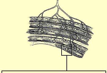
Link : Neuromuscular Junction ToxinsLink : Nicotinic receptor in the neuromuscular junctionLink: La tétrodotoxineLink: Neurotoxins
NICOTINIC ACETYLCHOLINE RECEPTORS
THE ROLE OF THE NEUROMUSCULAR JUNCTION IN MUSCLE CONTRACTION

To make a muscle contract, the acetylcholine produced in the presynaptic neuron of the neuromuscular junction must bind to the nicotinic receptors on the postsynaptic side. Each of these receptors consists of 5 subunits that form a pentagonal structure around a central channel.

Lien : Neurons: Animated Cellular & Molecular Concepts (click on 3. Ions and Ion Channels)


The nicotinic receptor is a channel (or ionotropic) receptor: the same protein both forms the transmembrane channel and binds the acetylcholine or one of its agonists, such as nicotine. When one of these substances binds to the receptor, the channel opens, allowing many sodium ions to enter the post-synaptic cell and a few potassium ions to leave it, thus depolarizing it.

There are two types of nicotinic receptors: N1, found in the autonomic nervous system, and N2, found at neuromuscular junctions. These receptors belong to a large family of channel receptors which include those for glycine, glutamate (NMDA and AMPA) and GABA (GABA-A receptors ).


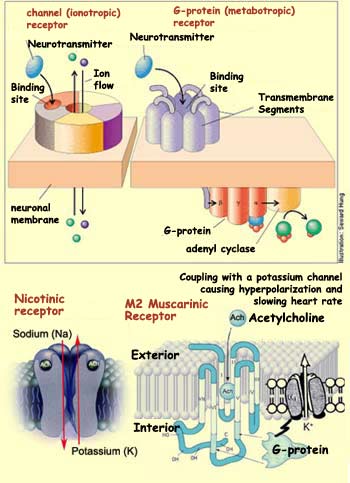
In addition to nicotinic receptors, there is another family of acetylcholine receptors, the muscarinic receptors. They belong to another large class of receptors called G-protein (or metabotropic) receptors. The receptors in this class (which includes dopamine receptors, for example), are totally separate from the ion channels. They exert their effects on these channels via a protein located on the cytoplasmic side of the cell, known as G-protein because it binds GTP. When a neurotransmitter binds to and activates a G-protein receptor, this receptor in turn activates this G-protein, which controls the opening of the physically separate ion channels, either directly or indirectly (via a second messenger). G-protein receptors therefore act more slowly than nicotinic receptors, where everything is centralized on the same protein complex.

Also, whereas nicotinic receptors are composed of five distinct peptides, the seven transmembrane domains of muscarinic receptors all come from a single protein that snakes its way back and forth across the membrane.

There are at least five different types of muscarinic receptors, all of which can be activated by muscarine, a molecule produced by a mushroom. M1 and M3 receptors, for example, activate phospholipase C, a second messenger that brings about depolarization by opening calcium channels while reducing the flow of potassium. In the brain, M1 receptors are found in the cortex and the central grey nuclei, while M3 receptors are found in the cerebellum. Both types of receptors are also involved in exocrine gland secretions.

The action mechanism for type M2 receptors is different. These receptors are coupled with a G-protein that inhibits adenyl cyclase. The reduction in the activity of this enzyme reduces the amount of the second messenger cyclical AMP, allowing the potassium channels to open and hyperpolarizing the cell. M2 receptors are also found not only in the central nervous system (cerebellum, central grey nuclei, and brainstem) but also in the heart.

  Presentations | Credits | Contact | Copyleft